

Lucas C. Parra @lcparra@qoto.org
- Website:
- https://parralab.org
neuroscience, machine learning, brain stimulation, brain signals
Joined Nov 2022
Lucas C. Parra
boosted

Difficulty with the preceding visual search affects brain activity in the following resting period
https://www.nature.com/articles/s41598-022-21624-3
Lucas C. Parra
boosted

YES. @jamesmharrison reminded me that "Inhibition is something that we DO".
But to tease apart dynamics of goal-directed activation amidst goal-directed suppression... especially when the goal may be unconscious or habitual in one instance, or skilled and volitional in another?
This new preprint may have the methodology needed to tease these things apart - "A mind-body interface alternates with effector-specific regions in motor cortex" 👇 https://www.biorxiv.org/content/10.1101/2022.10.26.513940v1
Lucas C. Parra
boosted

@neuroecology there seem to be various cross-posters, here is one: https://crossposter.masto.donte.com.br/
I'm not sure how it will really work given that often on twitter you're replying to an ongoing conversation ... I haven't tried it but post if you get to know it ...
Lucas C. Parra
boosted

Hi, this is Greta. You are welcome to follow me here, if you prefer planet earth to planet mars.
Lucas C. Parra
boosted

About 25 years ago, Crick and Koch wrote a very thought-provoking paper,
https://www.nature.com/articles/34584
It tries to answer the question, why do feedforward (FF) and feedback (FB) projections have such different properties in terms of terminating in different cortical layers and physiological impact — something that's ignored in many recurrent network models of cortex.
i.o.w.: Why do higher areas talk so differently to lower areas than lower areas talk to higher areas?
Lucas C. Parra
boosted

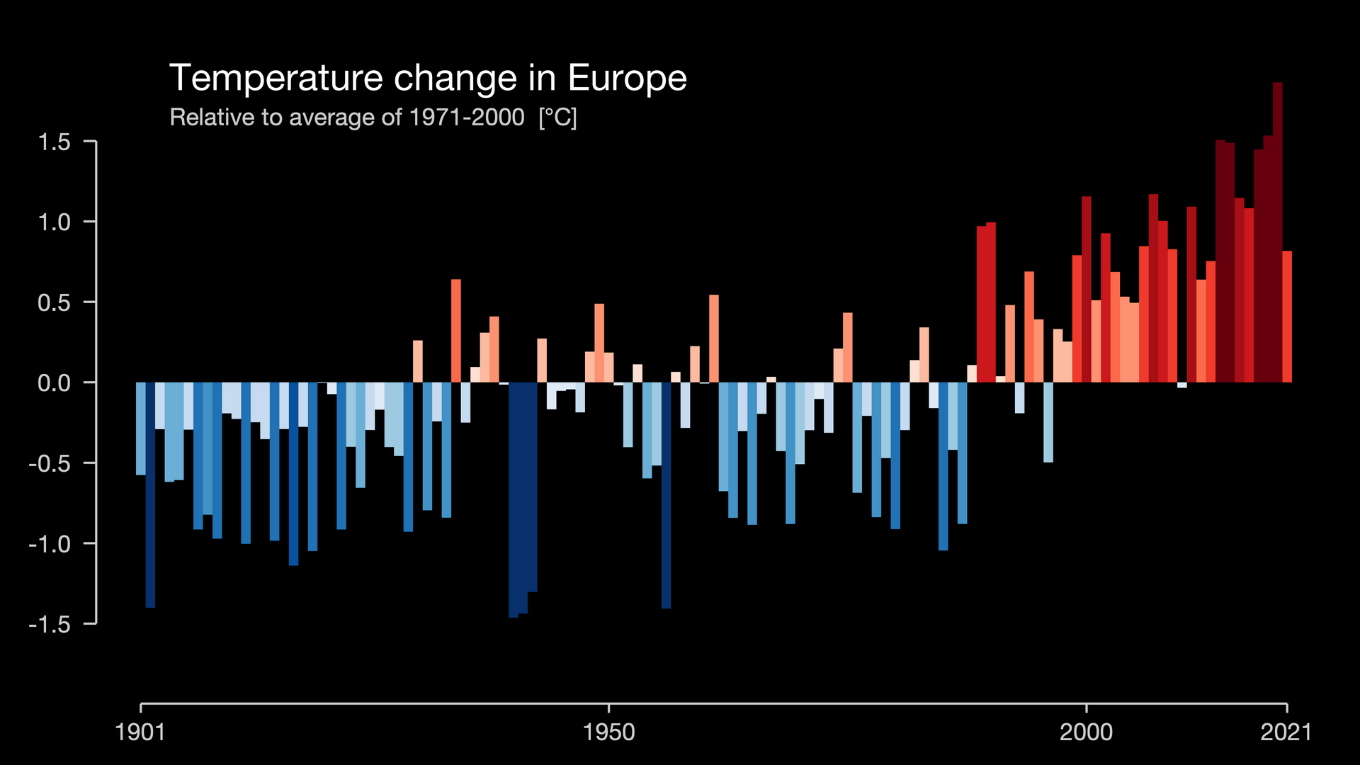
Average over all of Europe:
Temperature change, 1901-2021
https://showyourstripes.info/c/europe/all
Lucas C. Parra
boosted
Mastodon habits I'm trying to lock in, rather than revert to my Twitter habits:
1) use CWs liberally
2) when threading, set first post to "public" and the rest to "not listed"
3) don't forget the description text when posting images (had to work on that in Twitter too)
4) throw in hashtags like it was Tumblr or Instagram when you want to reach beyond your followers
5) pin and visit hashtags to find more people
6) boost a lot
Lucas C. Parra
boosted

For those of us who came from Twitter: here, clicking "favorite" is different from clicking "like" in Twitter. It just tells the writer "yeah, nice", without enhancing visibility. The only way to enhance visibility is to "boost" (basically, retweet). I suspect all this will change (different clients will have different algorithms, etc) but for now this is how it is...
@albertcardona I saw that too. I think he actually believes it.
Lucas C. Parra
boosted

Assessing pupil size as an index of activation of subcortical ascending arousal system nuclei during rest https://www.biorxiv.org/content/10.1101/2022.11.04.514984
Here is research in my lab: https://parralab.org
Will be posting about #AI #neuroscience #brain signals and brain stimulation.
For a history of posts see: https://twitter.com/lucas_c_parra
Lets get started ...
Narratives synchronize our hearts: https://www.pnas.org/doi/10.1073/pnas.2206199119

- Website:
- https://parralab.org
neuroscience, machine learning, brain stimulation, brain signals
Joined Nov 2022
