

Albert Cardona @albertcardona@qoto.org
- Personal website
- https://albert.rierol.net
How does the brain work? Someday, we'll figure it out.
Group Leader, MRC LMB, and Professor, University of Cambridge, UK.
#neuroscience #Drosophila #TrakEM2 #FijiSc #CATMAID #connectomics #vEM
Born at 335 ppm.
Brains, signal processing, software and entomology: there will be bugs.
Joined Oct 2022
Albert Cardona
boosted

Albert Cardona
boosted

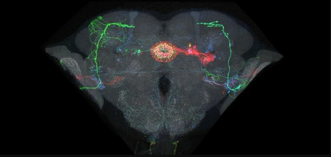
I've been sitting on this paper from @ScottishWaddell@twitter.com and @ManuPerisse@twitter.com because y'all know MB neuron names give me anxiety. But it's illustrated so well you don't need the names! Flies use both aversive and reward-coding DANs to judge relative value. https://doi.org/10.1016/j.cub.2022.08.058
Albert Cardona
boosted

This article nicely summarizes how NeuronBridge can be used to go from #connectome circuit to #fly strain by enabling access to large, open data sets. #drosophila #neuroscience #OpenScience
https://www.janelia.org/news/janelia-software-tools-power-fly-brain-research
Albert Cardona
boosted

RS-FISH was just released as open access article in @naturemethods
RS-FISH is a user-friendly software for accurate spot detection that is applicable to smFISH experiments, spatial transcriptomics, and spatial genomics. The approach enables fast spot detection in even very large volumetric datasets.
https://github.com/PreibischLab/RS-FISH
https://github.com/PreibischLab/RS-FISH-Spark
Albert Cardona
boosted

#introduction I'm a scientist at the MRC Laboratory of Molecular Biology in Cambridge, UK. Our group works on developing new #microscopes and #instruments for #StructuralBiology, especially #CryoEM. All the problems we work on are biological, but often turn into physics and then engineering and computation before being solved. A diversity of ideas, people, expertise and talents are the key to making it happen.
Need a good laugh?
Do you keep hearing about machine learning taking over the world "soon anytime now"?
This #deviantart entry of mine from 2009 has been labelled, presumably automatically, as "mature content". I kind of see why–not quite an adversarial attack, but yes quite the failure of excessive generalization.
It's a most innocent photograph: that of my favourite lunch, back from when I was at INI Zurich: mozzarella with cherry tomatoes, olive oil, salt, and ... bread.
Albert Cardona
boosted

#Postdoc position to investigate cGAS-STING-dependent signaling in drosophila. A three-year postdoc position starting in spring 2023 with Jean-Luc Imler and 🐘krin_meignin@drosophila.social at IBMC in Strasbourg (France). #immunity #drosophila #virus
Albert Cardona
boosted

Addiction
Don't post on Twitter.
The content and connections that you are investing there are supporting an abusive system.
Post that content here.
Bring your connections here.
There, you are small, and you are a commodity.
Here, you are important, and you are a person.
You know all this.
If you feel compelled to open Twitter, investigate that compulsion.
You've heard that algorithmic apps can cause addictive behavior.
Is that what you're doing?
And if so, shouldn't you stop?
Albert Cardona
boosted

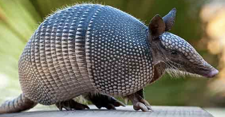
This is nuts: Using armadillos as a model system, this group has shown that #leprosy bacteria are able to reprogram differentiated hepatocytes into stem-like cells. This ultimately results in further cell differentiation and proliferation (which ultimately benefits the bacteria by increasing the host resources available). The paper highlights the possibilities for liver regeneration in medicine.
But we're here to be awed by this new parasite manipulation strategy.
Albert Cardona
boosted

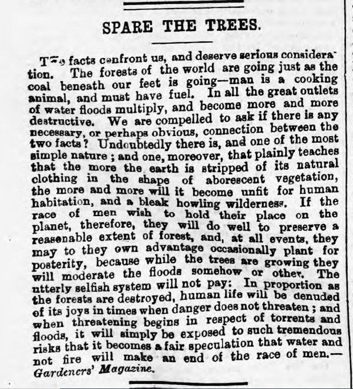
A warning from 143 years ago...
'The Aberystwyth Observer, 24th May 1879'
"...the more the earth is stripped of its natural clothing in the shape of arborescent vegetation, the more and more it will become unfit for human habitation, and a bleak howling wilderness."
How much as a species have we learnt?
https://newspapers.library.wales/view/3041425/3041431
Brought to our attention by @JasonEndfield Twitter
#Trees #ClimateAction #climatechange #ClimateCrisis #Cymru #Wales
Albert Cardona
boosted

@csdashm @albertcardona
To expand on the "cognitive fingerprint" comment, cognitive neuroscientists often treat the pattern of neural activations evoked by a stimulus or mental concept as a "fingerprint" for that concept.
For instance, I remember going to a talk by a social scientist where she showed similarities in the neural activity between two tasks: reading about companies interacting or reading about people interacting. In contrast, reading about objects interacting produced different neural responses. To see whether corporations are seen as people or objects, the specific areas activated are not important, but the similarity of the patterns is. The activation pattern is treated as a fingerprint: the grooves in the finger matter only in that they consistently identify the finger's owner.
Treating the neural activity as a fingerprint and inferring mental concepts is a big part of cognitive neuroscience. However, this can frustrate biologists who care about the specifics of the neural computations. What's more, in systems neuroscience, my impression is that behaviorism rules and it frowns upon inferences about the psychology of non-human animals. Thus the gap is even bigger!
Albert Cardona
boosted

@albertcardona I pretty much agree with you, but I think it depends also on what you're looking for.
To use a programming/computer hardware analogy, if you want to figure out an algorithm, fMRI is useless. If you want to know whether your computation is hitting RAM hard or requires a lot of floating point operations or is engaging the graphics card a lot (and whether shaders are used), it's pretty handy.
Rather than fMRI being a case of looking for the wallet where the light is, it's more like it's showing you where to put the light to look for wallets (if it's done well).
However, the number of overinterpreted fMRI studies does rather disappoint me. The inference "we didn't see a fMRI signal in this area so it's not important in this task" seems to happen far too often.
Then again, this is one of the biggest mistakes of scientists in every area: assuming that absence of evidence is evidence of absence when the tools and/or statistics were not specifically being deployed to address that exact question (i.e. putting bounds on how absent something is).
As a rough heuristic, I find it depressingly accurate to assume that a sentence starting with "This isn't significant, so" is one that will end with an unjustified conclusion.
Albert Cardona
boosted
@neurolili @albertcardona - Good points Lilli, but I do want to point out that the poor temporal resolution of old calcium indicators was one of the primary reasons why Janelia put so much effort into calcium indicators to get them to be fast, and also that even the slow ones at least have single-cell resolution. And speed is part of why GCaMP6 was so popular.
So I don't think that argument gets very far. (I didn't trust the slow calcium indicator signals all that much either.)
Albert Cardona
boosted

@neurolili @albertcardona as an in the weeds EM person like Albert, I feel like “fingerprints that clarify cognitive concepts” needs to be unpacked a bit for people who are mostly going after cellular-level mechanisms. One thing that’s hard is feeling like there are so many more details we know about drosophila or mouse visual cortex, and yet the feeling of “understanding” is still fleeting (albeit slowly coming into view in the fly, I think).
Albert Cardona
boosted
I'll note that I think part of the confusion is a cultural one. Having gone from a cognitive neuroscience fMRI/ECoG lab to a drosophila neuroscience lab, I feel there is a disconnect between the goals and methods between the two fields. The drosophila neuroscientists (unfairly) dismiss cognitive neuroscience as not rigorous enough, whereas the cognitive neuroscientists (unfairly) dismiss work on drosophila as they see flies as "too simple".
I think this reflects a larger gap that I've seen of neuroscientists approaching the brain from a cognitive versus biological angle and how this leads to them to pursuing different goals using different methods. Cognitive scientists are often looking at neuroscience for fingerprints that can clarify cognitive concepts, whereas biologists often look at neuroscience trying to understand natural computation and biological processes connecting to the rest of the body.
Albert Cardona
boosted
@albertcardona Thank you for this post! I do enjoy thinking about this topic. There's a lot to respond to here, so perhaps I can do it in pieces.
1) The BOLD signal (captured by fMRI) is only "somewhat correlated with neural activity"
It is correlated with neural activity, albeit there is a low-pass filter on top. It is most correlated with LFP signals in monkeys (Logothetis et al, 2001, https://www.nature.com/articles/35084005 ) and humans (Nir et al, 2007, https://www.cell.com/fulltext/S0960-9822(07)01635-1 ).
The BOLD signal is fairly slow, but you can deconvolve it and get temporal precision on the order of 0.5s or so (also subject to your sampling time). The lower temporal precision and indirect aspect of fMRI compared to ephysn is not so different from GCaMP sensors (especially earlier ones), which have been much less controversial.
For studying cognitive neuroscience, 0.5s can actually be really powerful, as you can set up much more complex tasks in humans and jitter stimulus presentation.
2) Each voxel is too large to see anything interesting
If decades of fMRI research have taught us anything, it's that brain areas do get engaged in a grouped enough fashion in order to measure it. Reward signals, for instance, are particularly salient in fMRI. Just that fact has been enough to get researchers excited about connecting economic rewards to their neural instantiation.
One of my favorite studies showing the power of fMRI, despite these bigger voxels, is a clever analysis showing how visual and language representations are right up against each other in the cortex (Popham et al, 2021, https://www.nature.com/articles/s41593-021-00921-6 ). In what other modality could you get such a result??
Besides, MRI technology is getting better and better. You can already get better spatial resolution if you image a specific brain region. There's also efforts to increase the spatial resolution overall by redesigning scanner components and improving signal processing, leading voxels that can image individual columns:
https://vcresearch.berkeley.edu/news/134-million-build-next-gen-mri-brain-scanner-uc-berkeley
3) Brain areas are plastic, so does it make sense to build a science around regions that generalizes across people?
The most general form of this argument makes neuroscience feel hopeless. Yes, if you wire visual signals to the auditory cortex of a marmoset, it might indeed process these. However, does that mean we should not study the development and fine structure of the visual cortex for processing vision? Surely not!
Plus, it's true that brain regions can be reorganized across people, but to me it amazing at how much is preserved! For instance, Wager et al (2013, https://www.nejm.org/doi/full/10.1056/nejmoa1204471 ) found a signature of pain that could predict a participant's pain even when trained on other subjects. This is not to downplay individual variability, I think all of neuroscience (across all methods and levels) needs to more fully grasp with this. That said, there are common patterns and they are already quite interesting on their own!
Albert Cardona
boosted

 🏳️🌈🖖🏽
🏳️🌈🖖🏽
I’m boldly going where I haven’t gone before, which is right here at this site. Apparently this is a “toot.” I would appreciate a follow!
Controversial take? I consider myself a neuroscientist, and I am not able to understand the usefulness of fMRI for cognitive neuroscience studies. (fRMI seems like a great tool to diagnose brain cancer, though.)
In fMRI, every voxel represents several cubic millimetres of brain tissue comprising millions of neurons; the temporal sampling is 2 seconds, when neurons fire action potentials in the ~10 millisecond range, and fast behavioural responses are in the ~300 millisecond range; and the signal measured is blood flow which is somewhat correlated with neural activity at those timescales.
fRMI studies in patients with chronically implanted electrodes (to detect the location of epileptic centres) seem to indicate that areas with low fRMI signal aren't necessarily "unimportant", on the contrary, a small percent of neurons in that area may be critical, yet their activity isn't captured in the fMRI signal as significant. Studies from Ueli Rutishauser and collaborators come to mind.
Then there's the issue of brain "areas". The study of the brain as made of compartments breaks down at close scrutiny. First, monitoring neural activity of the visual cortex in the absence of visual stimulus showed that neuron activity tracks body motion (Carsen Stringer et al. 2019 https://www.science.org/doi/abs/10.1126/science.aav7893 ); in other words multi-sensory integration is the norm. Second, high-functioning hydrocephalic cases present a greatly altered brain architecture with the grey and white matter occupying a tiny fraction of the overall volume. Third, accidents have revealed great plasticity in brain areas, with areas not being spatially stable but rather able to expand over adjacent areas that are less used because of e.g., a missing body part. Even complete absence of the entire cerebellum (cerebellar agenesis) can result in mild phenotypes (Yu et al. 2014 https://www.doi.org/10.1093/brain/awu239 ).
In other words, brain "areas" is not quite the useful abstraction we would want it to be. And therefore, fRMI imaging of blood flow changes over time across coarsely spatially and temporally sampled brains is, at best, too much of a low pass filter over the signal we'd be interested in monitoring.
Are fMRI studies a case of "there's more light here and therefore I look for my wallet here rather than overthere in the shadows where I can't see at all"? I understand that fRMI, and EEG, are all we have to study neural activity in the human brain, so there's a strong incentive to just go with that despite strong shortcomings. Am I missing something fundamental about fRMI?
The only studies using fMRI that make sense to me are longitudinal studies, where the same patient is imaged multiple times and comparisons are like to like, and have more to do with discovering structural issues related to e.g., ageing than assigning function to any subset of the brain, such as in Linda Geerligs' work (Geerligs et al. 2015 https://academic.oup.com/cercor/article-abstract/25/7/1987/462366 ). Are there any other kinds of fMRI studies that beyond doubt have contributed to our understanding of the human brain?
Albert Cardona
boosted

Another #introduction from someone considering leaving the dark side of the force ... #TwitterMigration
Trained in #cellbiology, I caught fire for all things EM pretty early on, brought #Austria's first dedicated facility using #cryoem to life in 2006, and started my own company Nexperion in 2013.
Now busy with lots of #SerialEM, #cryoem, #cryotomo, and a monitoring system for TEM developed by my company, named Sentinel.
Also: #archlinux, #sweden, ...
Albert Cardona
boosted

In case you were wondering:
@the_node@mstdn.science
@J_Cell_Sci@mstdn.science
@focalplane_jcs@mstdn.science

- Personal website
- https://albert.rierol.net
How does the brain work? Someday, we'll figure it out.
Group Leader, MRC LMB, and Professor, University of Cambridge, UK.
#neuroscience #Drosophila #TrakEM2 #FijiSc #CATMAID #connectomics #vEM
Born at 335 ppm.
Brains, signal processing, software and entomology: there will be bugs.
Joined Oct 2022
