

Albert Cardona @albertcardona@qoto.org
- Personal website
- https://albert.rierol.net
How does the brain work? Someday, we'll figure it out.
Group Leader, MRC LMB, and Professor, University of Cambridge, UK.
#neuroscience #Drosophila #TrakEM2 #FijiSc #CATMAID #connectomics #vEM
Born at 335 ppm.
Brains, signal processing, software and entomology: there will be bugs.
Joined Oct 2022
The #MRCLMB offers fully funded PhD studentships. Come join us!
Deadline: December 1st, 2022 (six days left).
Have a look at the projects listed for the Neurobiology Division (where Marta Zlatic, Greg Jefferis, Bill Schafer, Marco Tripodi, and many others, including myself).
List of projects: https://www2.mrc-lmb.cam.ac.uk/students/international-phd-programme/projects/
How to apply: https://www2.mrc-lmb.cam.ac.uk/students/international-phd-programme/how-to-apply/
One of the two first authors, Logan Thomas @lathomas42 is on mastodon, as is the senior author Wei Lee @darbly. Welcome! And what a spectacular paper on #cerebellum #connectomics. Those must be the prettiest Purkinje cell renderings since the century-old famous ones from Cajal. This time with synapses though!
#neuroscience
"Structured cerebellar connectivity supports resilient pattern separation" Nguyen, Thomas et al. in @darbly's lab https://www.nature.com/articles/s41586-022-05471-w
Spectacular work based on connectomic reconstruction from nanometre-resolution volume electron microscopy and computational modelling that contributes novel findings in cerebellar microcircuitry:
"both the input and output layers of the circuit exhibit redundant and selective connectivity motifs, which contrast with prevailing models. Numerical simulations suggest that these redundant, non-random connectivity motifs increase the resilience to noise at a negligible cost to the overall encoding capacity. This work reveals how neuronal network structure can support a trade-off between encoding capacity and redundancy, unveiling principles of biological network architecture with implications for the design of artificial neural networks."
#cerebellum #connectomics #neuroscience #science #vEM #volumeEM #NeuralBetwork
The impact of exercise on the brain was brought into focus, for me, by Stryker's lab work on approaches to recover from long-term vision deprivation through visual training while exercising physically ("stimulated by locomotion").
The underlying mechanism seems to be "the reduction produced by deprivation in thalamocortical excitation is compensated for during recovery by a corresponding reduction in the magnitude of inhibition."
Stryker wrote a review here:
"A neural circuit that controls cortical state, plasticity, and the gain of sensory responses in mouse" Stryker 2014 http://symposium.cshlp.org/content/79/1.short
Related, given that awake animals are often on the move:
"We have now studied the response properties of neurons in primary visual cortex of awake mice that were allowed to run on a freely rotating spherical treadmill with their heads fixed. Most neurons showed more than a doubling of visually evoked firing rate as the animal transitioned from standing still to running, without changes in spontaneous firing or stimulus selectivity."
"Modulation of visual responses by behavioral state in mouse visual cortex", Niell and Stryker 2010 https://www.sciencedirect.com/science/article/pii/S0896627310000590
Cortical area instability–experiments in monkeys
On the instability (plasticity?) of cortical areas, just got reminded of this classic work:
"Somatosensory cortical map changes following digit amputation in adult monkeys", Merzenich et al. 1984 https://onlinelibrary.wiley.com/doi/abs/10.1002/cne.902240408
“Exercise increases information content and affects long-term stability of hippocampal place codes” in mice. But the implications are tantalising.
Rechavi et al. 2022 https://doi.org/10.1016/j.celrep.2022.111695
This hard #DevBio finding stands out: runners showed increased neurogenesis in their hippocampus.
“To assess the levels of hippocampal neurogenesis, we injected mice with Bromodeoxyuridine (BrdU), a marker for proliferating cells. In accordance with previous results (van Praag et al., 1999b), we found a higher number of BrdU-positive cells and increased labeling density of doublecortin (DCX), a protein transiently expressed in newborn neurons, in the DG of runners (Figures 1B–1D). This increase in adult neurogenesis indicates that physical activity affected the physiology of hippocampal circuits in our experiments.”
Neuro-evo conference at HHMI Janelia on May 15-18, 2023. Join us for the third edition!
Application deadline: Jan 27 (11:59 p.m. EST) 2023.
"Historically, with the study of the most convenient animal models —from the giant axon of the squid and the lobster's stomatogastric circuits to Aplysia's synapses and C. elegans' circuits — neuroscientists revealed some of the operating principles of the nervous system, which were then found to apply broadly across phyla. The third instalment of this meeting will once again bring together neuroscientists working on a broad diversity of animal models in an effort to compare circuits across phyla as a means to crack their function."
#NeuroEvo #Janelia #HHMI #conference #science #academia #neuroscience #DevBio #connectomics #connectome
Apropos #fMRI and comparing brain areas across subjects: variations of 3.5x for brain cortical area V1-3 across 118 human subjects.
"Variability of the Surface Area of the V1, V2, and V3 Maps in a Large Sample of Human Observers"
Benson et al. 2022 https://www.jneurosci.org/content/42/46/8629
HT: @Whatishealth
I think this new paper in J.Nsci is very important.https://www.jneurosci.org/content/42/46/8629 showing that V1-V3 in human across 118 subjects v...
“The natural history of Madagascar”, a new book including all those teeny tiny frogs and lizards—chameleons, in particular—with so much potential for whole-brain #connectomics, like the chameleons of the genus #Brookesia https://en.m.wikipedia.org/wiki/Brookesia
#herpetology #lizards #frogs #Madagascar #nature #neuroscience
HOT OFF THE PRESS: The New Natural History of Madagascar! At >2000 pages over two volumes, with >600 contributing authors, it sets a new benc...
When a GABAergic neuron synapses onto the axon of a sensory neuron, what happens?
Turns out, in the proprioceptors of the locust leg (chordotonal somatosensory neurons), "All the available evidence indicates that the inputs that occur during walking are depolarizing, inhibitory, synaptic inputs."
Depolarizing, inhibitory. How's that possible? Depolarizing because of where the resting potential of the axon is (which is different than the dendrite's), and the particularities of the GABA receptor on the receiving axons. Inhibitory because the depolarization is subthreshold, reducing the membrane voltage potential and, therefore, reducing neurotransmitter release when a spike arrives.
What is this good for? Implementing an efference copy signal: "The rhythmic depolarization at different phases of the step cycle in different sensory neurons may represent a predictive action of the CNS when generating a motor pattern." That is, predicting when the proprioceptor will fire, and by how much, as a function of the self-induced motion. So this signal is subtracted in order to be able to measure (and react to appropriately) any unexpected signal (such as the leg skidding or hitting a rock).
Wolf and Burrows, 1995. "Proprioceptive sensory neurons of a locust leg receive rhythmic presynpatic inhibition during walking" https://www.jneurosci.org/content/15/8/5623.short
Burrows and Matheson, 1994. "A presynaptic gain control mechanism among sensory neurons of a locust leg proprioceptor" https://www.jneurosci.org/content/14/1/272.short
Controversial take? I consider myself a neuroscientist, and I am not able to understand the usefulness of fMRI for cognitive neuroscience studies. (fRMI seems like a great tool to diagnose brain cancer, though.)
In fMRI, every voxel represents several cubic millimetres of brain tissue comprising millions of neurons; the temporal sampling is 2 seconds, when neurons fire action potentials in the ~10 millisecond range, and fast behavioural responses are in the ~300 millisecond range; and the signal measured is blood flow which is somewhat correlated with neural activity at those timescales.
fRMI studies in patients with chronically implanted electrodes (to detect the location of epileptic centres) seem to indicate that areas with low fRMI signal aren't necessarily "unimportant", on the contrary, a small percent of neurons in that area may be critical, yet their activity isn't captured in the fMRI signal as significant. Studies from Ueli Rutishauser and collaborators come to mind.
Then there's the issue of brain "areas". The study of the brain as made of compartments breaks down at close scrutiny. First, monitoring neural activity of the visual cortex in the absence of visual stimulus showed that neuron activity tracks body motion (Carsen Stringer et al. 2019 https://www.science.org/doi/abs/10.1126/science.aav7893 ); in other words multi-sensory integration is the norm. Second, high-functioning hydrocephalic cases present a greatly altered brain architecture with the grey and white matter occupying a tiny fraction of the overall volume. Third, accidents have revealed great plasticity in brain areas, with areas not being spatially stable but rather able to expand over adjacent areas that are less used because of e.g., a missing body part. Even complete absence of the entire cerebellum (cerebellar agenesis) can result in mild phenotypes (Yu et al. 2014 https://www.doi.org/10.1093/brain/awu239 ).
In other words, brain "areas" is not quite the useful abstraction we would want it to be. And therefore, fRMI imaging of blood flow changes over time across coarsely spatially and temporally sampled brains is, at best, too much of a low pass filter over the signal we'd be interested in monitoring.
Are fMRI studies a case of "there's more light here and therefore I look for my wallet here rather than overthere in the shadows where I can't see at all"? I understand that fRMI, and EEG, are all we have to study neural activity in the human brain, so there's a strong incentive to just go with that despite strong shortcomings. Am I missing something fundamental about fRMI?
The only studies using fMRI that make sense to me are longitudinal studies, where the same patient is imaged multiple times and comparisons are like to like, and have more to do with discovering structural issues related to e.g., ageing than assigning function to any subset of the brain, such as in Linda Geerligs' work (Geerligs et al. 2015 https://academic.oup.com/cercor/article-abstract/25/7/1987/462366 ). Are there any other kinds of fMRI studies that beyond doubt have contributed to our understanding of the human brain?
Job ad: Group Leader position in computational neurobiology https://cbd.sites.vib.be/en/join-us#/job-description/51111
Place: VIB-KU Leuven Center for Brain & Disease Research, Belgium.
Notice there are many other advertised job positions at that same research center, including lab tech, postdocs, #electronmicroscopy experts (#vEM), #proteomics specialists, administrative, and more: https://cbd.sites.vib.be/en/join-us#/job-list
For neuroscientists attending #sfn22: don’t miss the poster by Mitya Chklovskii’s group describing the completion status of the whole brain #connectome of the fairy wasp #Megaphragma, of expected completion early in 2022. Find the poster tomorrow Monday morning, number 328.16 / YY35.
Mitya kindly shared the poster image publicly elsewhere.
This tiny #wasp is famous for being the size of a large paramecium (a unicellular organism) and for enucleating the vast majority of its central neurons while pupating. The adult has less that 10,000 neurons in its central brain yet it isn’t missing any organ or body part. See the paper that jumpstarted this effort:
Polilov AA. The smallest insects evolve anucleate neurons. Arthropod structure & development. 2012 Jan 1;41(1):29-34. https://www.sciencedirect.com/science/article/pii/S1467803911000946
As an electron microscopist, every time I image a partial volume of the brain, I notice how most inputs and outputs of the neurons within originate in neurons outside the imaged volume. #microscopy
As a neuroscientist, when I reconstruct a neuron from #vEM I see that its inputs are collected from one or more brain areas, and its outputs target one or more other areas. There are local neurons but these are the exception. #neuroscience
As a programmer, I see that a software function can't be studied or understood in isolation, unless it''s a pure function, which is rare. Even functional programming languages such as #clojure or even #haskell require a fair share of non-pure functions in order to interact with the broader world.
As a developmental biologist, I see how one neuron is made after another in precise spatio-temporal patterns essential to assembling the correct circuit architecture. The study of any one neuron only makes sense within the context of the other neurons, and glia, and blood vessels, and more. #DevBio
As an evolutionary biologist, I notice how the fitness of an individual depends not on this or that neuron, but rather, on the effect, recursively, of one neuron on many other neurons. Brain modules are not pure, not enclosed, but mere shorthand to refer to broad chunks of an indivisible whole. Crutches for our present inability to grasp a collective so large and complex.
Yet the unit of selection is not even the whole brain, or the whole individual organism, but the population with its many relationships across its individuals, and even beyond, the interrelated collective of species that we call an ecosystem. Ecosystems are also under selection pressure, and they change. We are changing ours now. #evolution
Whole brains, whole individuals, whole ecosystems. Ultimately, the whole planet, as eloquently articulated by Lynn Margulis and Dorion Sagain in their 1989 book "Biospheres from Earth to Space". But I am satisfied, in the short term, with the study of the brain as a whole.
Alex Gomez-Marin reviews @PessoaBrain ’s latest book, “The entangled brain”:
“…offers a way to construe the brain as a fully integrated organ, a framework that “while not rare, is also not mainstream among neuroscientists.” A “divide-and-conquer strategy” has produced ever more refined brain maps, he argues, and subsequent leaps from structure to function. However, not only are anatomical brain areas far from simply located units of cognition but, as the subtitle of the book makes explicit, perception, cognition, and emotion are also interweaved.”
“In turn, proper anatomy calls for embryology. And, as tackled later in the book, evolution also informs brain organization. Disciplines, we learn, are entangled too.”
Science Transcending reductionism in neuroscience https://www.science.org/doi/10.1126/science.ade8689#.Y22_k3_qW1U.twitter
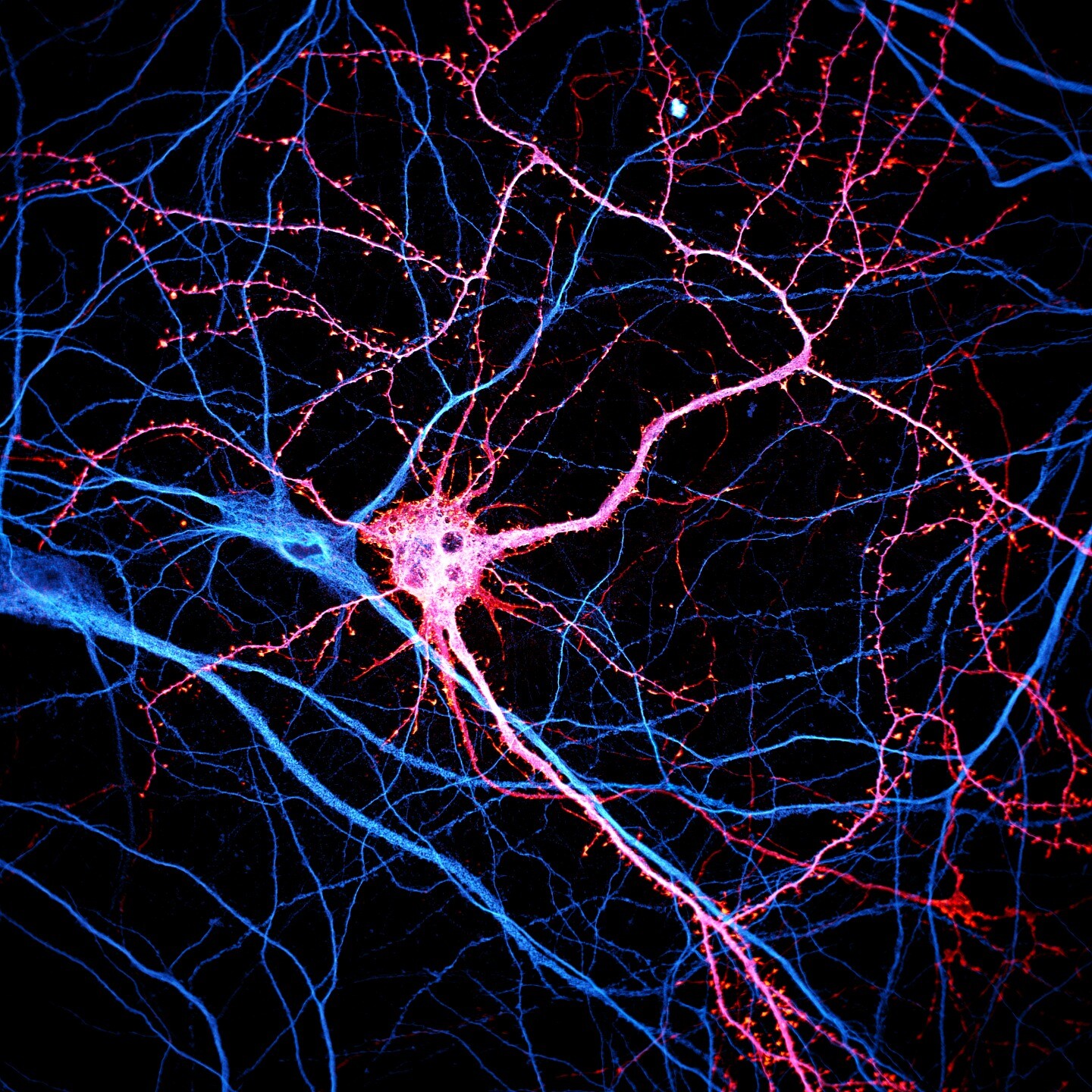
Beautiful fluorescent capture of a neuron by @chrislet@mas.to lab.
#neuroscience
Let's start with a pic from one of our microscopes! Neuron expressing tagged actin (orange) and stained for map2 (blue), spinning disk confocal ima...
Seminar starting now over zoom! Featuring @NicoleCRust #neuroscience
Looking forward to sharing this work - tomorrow (Nov 8, Tuesday, 8a EST / 3p IST) at BIU in Isreal, from Philly. The transformation from seeing to...
"Banburismus": find out what it is, how it relates to the breaking of the Enigma code in WWII, where the name comes from, and what does it have to do with #neuroscience:
@lakens Thank you for sharing this chapter! I didn't know the word "likelihoodist" and now I know I am one. Btw the brain is a likelihoodist too! I...
Casey Schneider—Mizell studies the #mouse cerebral #cortex with #vEM #connectomics at the Allen Brain Institute, and develops software for mapping and analyzing #neuronal #circuits in very large image volumes with nanometer resolution measuring over a cubic millimeter.
#neuroscience
Time for an #introduction. Nervous systems — yours, mine, those of mice, fish, and insects and worms — are made up of populations of different kind...
“Facemap: a framework for modeling neural activity based on orofacial tracking” Syeda et al. 2022 #neuroscience #biorxiv
New preprint by Atika Syeda and others in Carsen Stringer’s group. In Carsen’s words: Neural activity across cortex is high-dimensional and ... com...

- Personal website
- https://albert.rierol.net
How does the brain work? Someday, we'll figure it out.
Group Leader, MRC LMB, and Professor, University of Cambridge, UK.
#neuroscience #Drosophila #TrakEM2 #FijiSc #CATMAID #connectomics #vEM
Born at 335 ppm.
Brains, signal processing, software and entomology: there will be bugs.
Joined Oct 2022
