

DGE young microscopists @ydge@qoto.org
- Website
- www.ydge.de
- https://twitter.com/_ydge
- linkedin.com/company/dge-young-microscopists
Connecting students and postdocs from the German Society for Electron Microscopy (DGE) and beyond
Joined Nov 2022
Calling all budding #microscopists! 👩🔬 👨🔬 Don't miss our virtual International #Microscopy #studentmixer on May 7th, 2023 at 6 PM UTC via Zoom. Join us for relaxed conversations, online games, and a chance to win one of three raffle draw prizes sponsored by Thermo Fisher, MAS, and MSA. Register at https://forms.gle/MK4e7oPhYmuisGTw7. See you there! 🔬
It's the second day of #MC2023. Especially as an early career scientist, you should not miss our #yDGE #symposium later this evening at 18:15 (6:15 pm) in the room vanadium at darmstadtium.
Ph.D. student and expert talks 🥳
With the Microscopy Conference 2023 approaching, we would like to share ten tips for folks who are going to a conference for the first time.
1) Preparation:
1. Don't worry: A conference can be overwhelming, lots of people, large venue, many parallel sessions. But just observe and experience :)
2. Search the conference agenda for possibilities to connect to other students at Young Scientists events. These can be called student mixers and/or can be organized by the ‘youth association’ of the conference’s parent organization (eg. DGE/yDGE).
3. Look through the program and find out which sessions you definitely don't want to miss.
4. Dress code? It's a scientist's event, but it could be wise to look a bit more professional during your own presentation (sorts of 'smart casual', suits or costumes not needed)
a. If the question really gives you no peace: Check out the photos from past conferences in your field. You can usually get a good feeling on what the ‚dress code consensus‘ is.
5. You will usually walk around a lot: wear comfortable shoes
6. Can happen: venue is overly air-conditioned -> be prepared and bring a sweater or jacket
2) During the conference:
1. Find out where you will get food+beverages (lunchtime lectures, coffee breaks, shops around the venue)
2. Try to connect known names to faces.
3. Poster sessions are especially useful to make contact, and presenters are usually happy if you reach out to them. Simply saying that you're new to the field and asking if they can briefly say what their work is about is OK!
4. Remember: You are not the only first-timer at the conference!
🌐 📝
In addition, we would like to advertise our yDGE Symposium:
It will take place on 28/02/2023 from 18:15 until 20:15 in the Vanadium room.
Right after, we will also host a Social Networking Session. We are very much looking forward to connect with you in person in Darmstadt.
Do you have additional tips for conference newcomers? Please share them below!
DGE young microscopists
boosted

Want to work at the intersection of morphometry and representation learning for bioimage analysis?
We have TWO open postdoctoral positions! Each comes with its own challenging biological question, unique dataset, and exceptional team of collaborators!
Details below👇
DGE young microscopists
boosted

The identity of extra densities in #cryoEM maps of #amyloids from brains remains unknown, and a major focus of research. Just because a model fits in these fuzzy densities does not mean much, and writing speculative papers is going to be of little help.
https://www.biorxiv.org/content/10.1101/2023.02.01.526613v1
DGE young microscopists
boosted

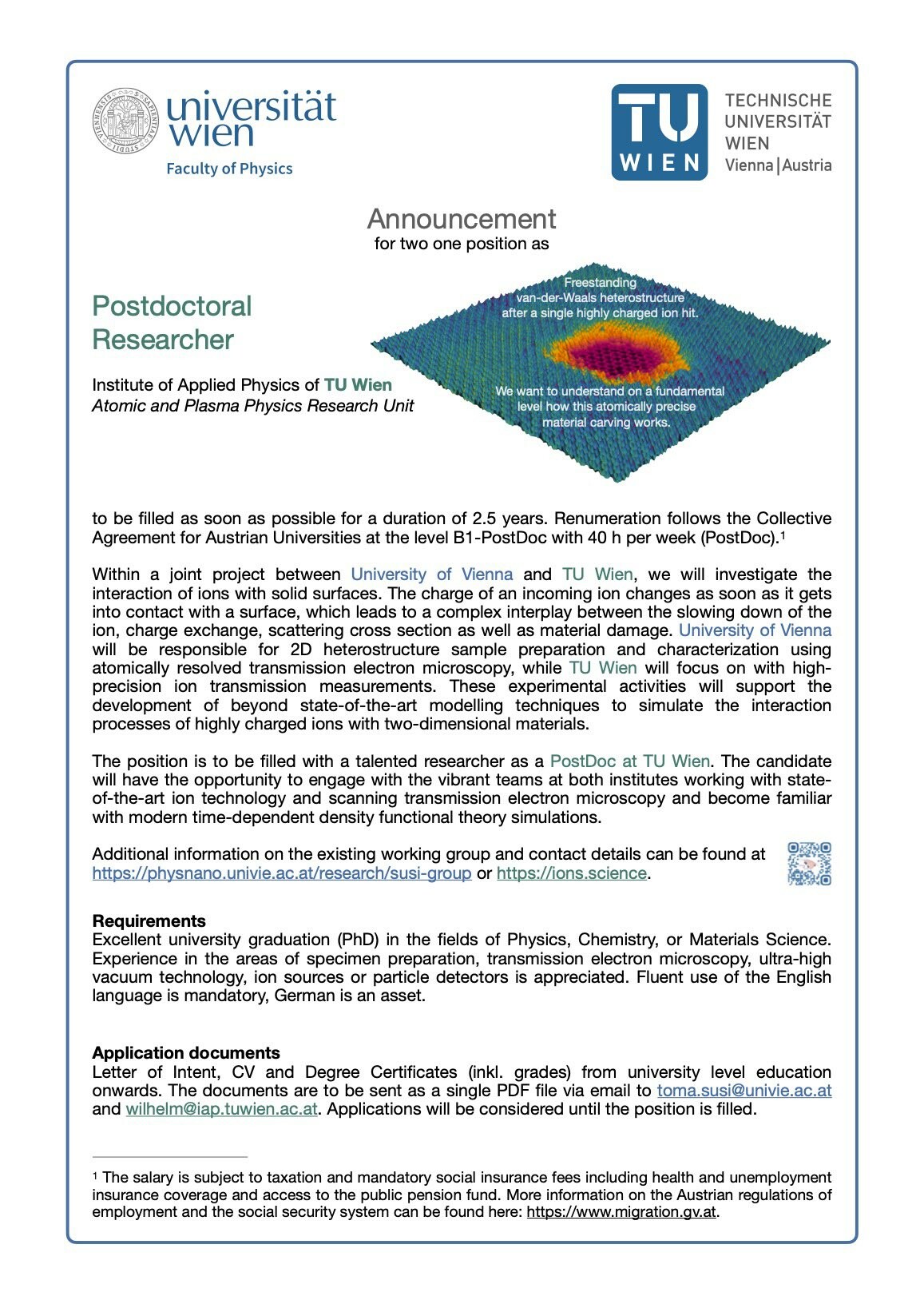
A collaborator of mine at TU Wien is looking for a postdoctoral research to join our joint project – fun with ions and and electron microscopy in 2D materials!
DGE young microscopists
boosted

Martha Montané-Romero et al. explore novel roles for #PLPP3 in #pluripotency exit and endodermal #differentiation in mouse embryonic stem cells:
#Lipids #StemCells #YAP1 #HIPPO #DevBio #Development #Science #BiologyOpen #Biology #AcademicMastodon
DGE young microscopists
boosted

a beautiful highlight of the microscopy by the Prevedel Lab: imaging 1.5mm into the brain #3Pmicroscopy #adaptiveoptics
DGE young microscopists
boosted

I have this silly question about #imagesegmentation. So is there any advantage to using different software for image segmentation? I see #ImageJ, #CellProfiler, #Napari and #Python all can do this, but if the underlying algorithm is the same, then why not just stick with ImageJ?
DGE young microscopists
boosted

#LaminarShear pushes VE-PTP (How?🤓) to downstream pole of #EndothelialCell, followed by endocytosis->⏫p-Tie2
Potentially explain how #DisturbedFlow⏫#VascularPermeability & #Atherosclerosis
Inhibition of VE-PTP by AKB-9785 fortifies #EndothelialBarrier & reduces Atheromas at atheroprone regions (aortic arch inner curvature, arch+intercostal bifurcations)🐭
Nice comparison of anti-leakage strategies
VE-PTP inhibition
Angpt1 activation of Tie2
Angpt2 inhibition
Dr. Donald McDonald & Dietmar Vestweber labs EMBO Mol Med 2023
https://www.embopress.org/doi/
DGE young microscopists
boosted

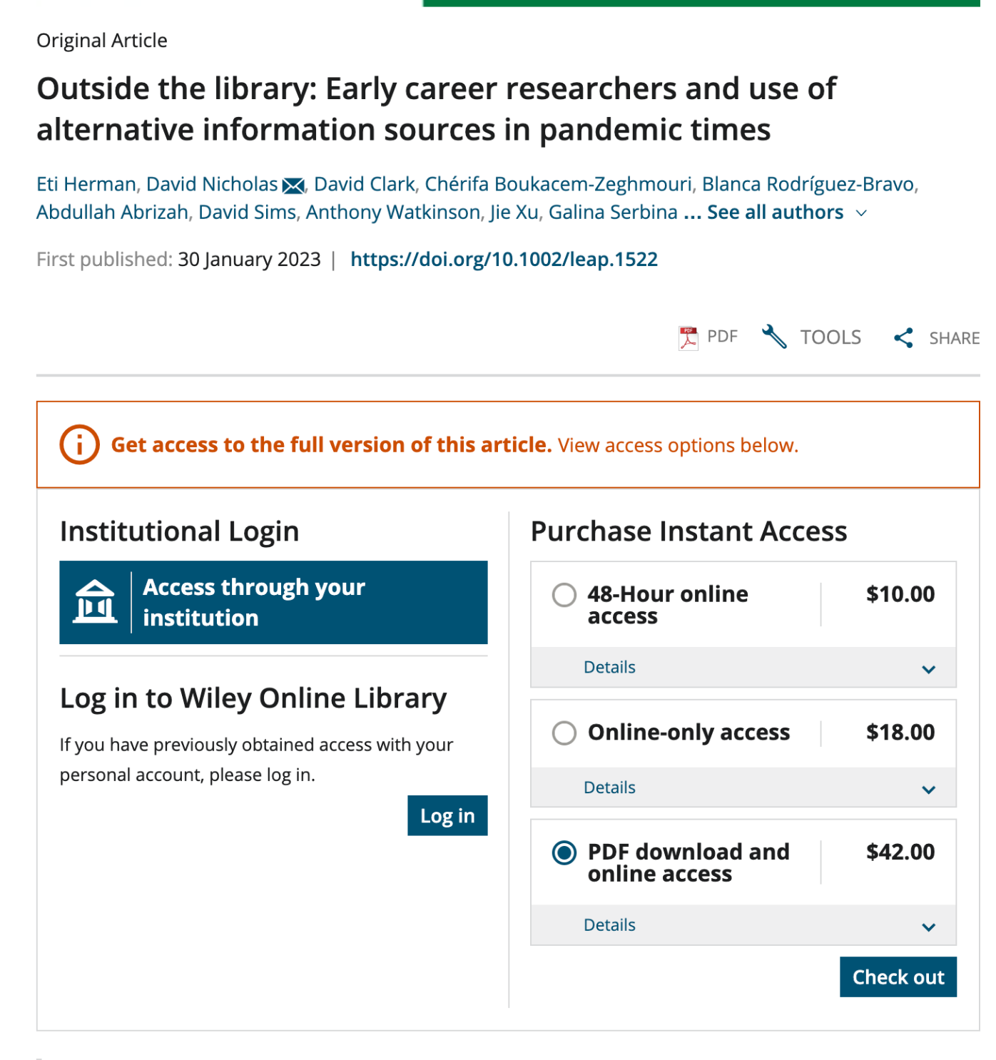
Ich soll $42 bezahlen, damit ich ein Paper herunterladen darf, in dem untersucht wird, warum junge Wissenschaftler Papers lieber im Netz raubkopieren, anstatt dafür zu bezahlen.
DGE young microscopists
boosted

RT: @svi_huygens@twitter.com
#microscopists, want to get a better overview of microscopy image #FileFormats, bit depth, scaling, image #metadata, dimensions, and more?
Join our webinar Feb 23 and explore what formats e.g. #OMETIFF:
DGE young microscopists
boosted

On this #FluorescenceFriday we are mesmerised by the process of cilium disassembly.
Find out more about the role of CCDC66 in regulating cilium length and signalling in the @J_Cell_Sci paper from Ezgi Odabasi, Deniz Conkar, @CytoLab & colleagues.
Also check out the tweetorial from Ezgi Odaba, which includes a movie showing assembly!
DGE young microscopists
boosted

RT @HoogendoornLab
We are recruiting a PhD student and/or postdoc! Are you interested in chemical biology, cellular signaling and the primary cilium? Look no further and come join us in beautiful Geneva 🇨🇭: https://www.hoogendoornlab.org/joinus Please RT!
@ERC_Research @unige_en @sciences_UNIGE
Time flies by. ⌛ One year ago, the DGE Young Microscopists (yDGE) were founded. In the first year, we set up a website www.ydge.de, 🌐 hosted our first social events and planned the first yDGE conference contribution 👔 (check out https://www.microscopy-conference.de/2/programme/scientific-programme WS 3).
#mc2023 #microscopyconference #microscopy #microscope #electronmicroscopy #electronmicroscope #science #imaging
DGE young microscopists
boosted

Paper alert 🚨😊 (at #science & electron #microscopy folks)
After our riCOM paper(https://doi.org/10.1017/S1431927622000617) we now published another #4D-STEM live reconstruction method. Here we use a Neural Network to reconstruct high-resolution phase images. It's all #opensource if u want to give it a go. 🙂
https://academic.oup.com/mam/advance-article/doi/10.1093/micmic/ozac002/6985579
- Website
- www.ydge.de
- https://twitter.com/_ydge
- linkedin.com/company/dge-young-microscopists
Connecting students and postdocs from the German Society for Electron Microscopy (DGE) and beyond
Joined Nov 2022
