

Joseph P. @tonic@qoto.org
I'm playing my part to support #biomanufacturing #biofabrication #biofoundry(ies) and bear witness to the birth of an industry. Working with 2 novel compounds to grow bone, cartilage & other types of cells. turns out it also works for growing hearts @organamet
Joined Nov 2022
@explainpaper The Uprising of Mitochondrial DNA Biomarker in Cancer
Authors : Siti Zulaikha Nashwa Mohd Khair, Siti Muslihah Abd Radzak,and Abdul Aziz Mohamed Yusoff
The Uprising of Mitochondrial DNA Biomarker in Cancer
Advances in predictive #diagnostic and #precisionmedicine, can lead to powerful discoveries and treatments for patients. #Cancer cells acquire functional capabilities to survive, proliferate, and circulate due to an enabling characteristic called genomic instability. Genomic maintenance systems have the ability to spot and repair any #DNA defects, while cancer cells increase the rates of mutation that orchestrate tumorigenesis. Chromosomal instability (CIN) is one of the most frequent changes observed in cancer cells, which often results from aberrations in chromosome structures and numbers. The second section of the paper focuses on #biomarkers, which are substances, structures, or processes that can influence or predict the incidence and outcome of a disease. There are three classifications of biomarkers: exposure, effect, and susceptibility. Biomarkers of exposure measure exogenous chemicals or their metabolites within an organism, while biomarkers of effect measure alterations of endogenous factors caused by exposure to an exogenous agent. Biomarkers of susceptibility measure genetic polymorphism predisposition of individuals and their external multifactorial influencers. Surrogate endpoints are often used to substitute clinical endpoints, and biomarkers can be used as a screening tool for an early indicator of #malignancy-risk development. They can also be used as diagnostic aids and prognostic biomarkers, as well as predictive biomarkers to identify the sensitivity and/or resistance of cancer patients towards specific agents or #medical product exposure.
The communication between the nucleus and mitochondria of a cell is known as intergenomic #crosstalk and it is bidirectional, meaning it can go both ways. It is important for regulating energy metabolism and tumor suppression. The communication is achieved by pathways such as anterograde #signaling and retrograde signaling. Anterograde signaling is when the nucleus controls gene transcription and cytoplasmic mRNA translation in response to external signals. Retrograde signaling is when mitochondrial dysfunction or loss of mitochondrial #membrane potential triggers communication with the nuclear genetic compartment. This communication is important for #homeostasis adaptation and can detect any nuclear damage or nuclear stress.
#Mitochondria are organelles found in cells that are responsible for producing energy. They are believed to have originated from a #singlecell-ed organism and are made up of two membranes. They contain their own genetic material, called #mtDNA, which is made up of 16569 nucleotide base pairs. mtDNA mutations can lead to mitochondrial dysfunction, which can cause #onco-genic events, such as #tumor cell reprogramming and metabolic shifts. #Mitoepigenetics is the study of how #epigenetics mechanisms regulate mtDNA transcription and replication, and it is believed to be involved in cancer progression.
The interconnection between #carcinogenesis (the development of cancer) and mitochondria (the energy-producing organelles in cells) was first proposed in 1973. Since then, there have been many studies done on this topic, using DNA scanning technologies to detect mutations and deletions. Mitochondrial DNA (mtDNA) is beneficial for carcinogenic studies because it consists of 37 genes with no introns, meaning most mutations will occur in coding regions. Additionally, mtDNA has a small size, is easy to extract, has no genetic rearrangements, and has fast mutation rates, which makes it useful for molecular research. It also has a high copy number, meaning only minimal #tissue samples are needed for analysis. Large-scale deletions are commonly known to be responsible for mitochondrial diseases, and are thought to be the cause of various diseases and cancers.
Two types of mtDNA deletions, 3.4 kb and 4977 bp are associated with various types of cancer. The 3.4 kb deletion was patented by Parr et al. [97] and is used to detect cancer in individuals. It is also used to determine different #prostate tissue types, either benign, malignant, or proximal to malignant [100]. The 4977 bp deletion is primarily associated with #aging and is a common deletion with missing mtDNA nucleotide sequences starting at 8470 to 13447 np [106]. It has been studied in various types of cancer, such as #breastcancer, #colorectal, gastric, hepatocellular, and #brain tumors, and is thought to be associated with external environmental factors, genetic predisposition, and ethnicity.
The text is discussing different types of deletions in mtDNA (mitochondrial DNA) that are associated with cancer. The 5.1.3 section is talking about the 3895 bp deletion, which was first observed in 1991 in two patients with progressive external #ophthalmoplegia. It was then found to be 10 times less frequent than the 4977 bp deletion. A study involving 104 age-matched subjects showed that the 3895 bp deletion was more frequent in those with usually sun-exposed skin and nonmelanoma skin cancer. The 4576 bp deletion was then discussed, which was found to be an indicator for breast cancer in a study involving 39 breast cancer patients. The 4576 bp deletion was not found in 23 normal patients without breast cancer. The text then moves on to discuss mtDNA copy number, which is the amount of mtDNA in each cell. It is suggested that mtDNA copy number changes may lead to mitochondrial instability and regulate energy metabolism, which can initiate #tumorigenesis. Studies have also shown that mtDNA copy number changes can be used as a predictive biomarker for #chemotherapy response.
Cell-free mtDNA (cf-mtDNA) is a type of mitochondrial DNA that is released into the #blood circulation due to disruption of the normal mitochondrial life cycle. It is believed to activate the Toll-like receptor 9 (TLR9) pathway, which can cause #inflammation and potentially lead to cancer. It has been used to diagnose cancer and sepsis, and as a biomarker for metabolic syndrome and predicting the risk of future diabetes. It is also being studied as a #noninvasive liquid #biopsy for cancer, as higher levels of cf-mtDNA have been found in cancer patients compared to healthy controls. Research is being conducted to find the potential link between cf-mtDNA and various cancers, as it is a preferable biomarker due to its higher mtDNA copy number, simpler structure, and shorter length.
Mitochondrial Microsatellite Instability (#mtMSI) is a type of genetic mutation that occurs in the mitochondrial genome. It is caused by short tandem repeats (mononucleotide or dinucleotide) of 1 to 6 base pairs that are scattered throughout the mitochondrial genome. These variations can lead to frameshift mutations, which can be caused by DNA polymerase γ, an enzyme that is responsible for oxidative damage. Mammalian mitochondria also have an inefficient mismatch repair system, which can lead to mtMSI formation. The most commonly reported mtMSI is located in the D-loop region, which is a mutational hotspot in primary tumors. It is a highly polymorphic homopolymeric C stretch, which is involved in R-loop formation, a stable RNA-DNA hybrid that triggers mtDNA replication. D310 alteration has been suggested as a new cancer detection tool and a potential early premalignant cancer marker. Another potential marker is D16184, which is located in the hypervariable region I and is involved in mtDNA biogenesis. Studies have reported the presence of D16184 in various cancer types, such as gastric and endometrial carcinoma. Somatic mtDNA alterations have also been correlated to cancer, with evidence showing that mtDNA changes can contribute to the development or progression of cancer. One example of a somatic mtDNA alteration is A12308G, which is located in the variable loop next to the anticodon stem of tRNA Leu (CUN). This alteration has been suggested as a potential diagnostic tool for #colorectalcancer and as a risk factor for prostate and #renalcancer. A10398G has also been studied in relation to cancer, although the results have been conflicting.
Mitochondrial biomarkers are molecules that can be used to detect cancer in its early stages. A commercial kit (PCMT) has been developed to help with this detection. However, even if cancer is detected early, it can still be difficult to treat if the symptoms have not yet developed. Therefore, researchers are looking into gene therapy and other mitochondrial interventions as potential treatments for cancer. They can use current advancements in vitro mitochondrial intervention to identify the pathogenicity and therapeutic potential of a particular mtDNA mutation. One method proposed is to transfer artificial healthy mitochondria to remove damaged mtDNA without genetic manipulation. Other studies have looked at the levels of mtDNA biomarkers in cancerous and non-cancerous samples, as well as the levels of mtDNA methylation and mtRNA in cancerous tissues.
@Renalcancer@a.gup.pe
@Brain@a.gup.pe
@Breastcancer@a.gup.pe
Just wanted to add this one to @explainpaper hope this works
The Therapeutic Potential of Exogenous Adenosine Triphosphate (ATP) for Cartilage Tissue Engineering
authors : Jenna Usprech , Gavin Chu , Renata Giardini-Rosa , Kathleen Martin , and Stephen D. Waldman
#Articular #cartilage, which is a type of #tissue found in #joints, allows for nearly frictionless motion and can absorb large loads. Unfortunately, when it is damaged, it cannot repair itself. #Tissueengineering is a promising approach to repair the damage, but it falls short of creating functional tissue. This is because the tissue-engineered constructs do not have the same mechanical properties as native articular cartilage, which is due to the insufficient accumulation of #extracellular matrix components. To address this, researchers have been exploring the use of adenosine triphosphate (#ATP) to directly harness the underlying mechanotransduction pathways responsible. ATP is a molecule that is released as a result of mechanical stimulation and acts as an autocrine/paracrine signaling #molecule. It acts on P2 receptors on the #plasma #membrane to promote extracellular matrix #synthesis. However, high doses of ATP can lead to an increase in matrix #metalloproteinase 13 (MMP-13) activity and extracellular inorganic pyrophosphate (ePPi) accumulation, which can lead to undesirable effects such as #mineralization of articular cartilage. Therefore, the purpose of this study is to identify the mechanism of ATP-mediated #catabolism and to determine a therapeutic dose range to maximize the #anabolic effect.
Materials & Methods
Cell Isolation: This is the process of separating cells from a tissue sample. It is usually done using #enzymes to break down the tissue and then filtering the cells out.
3-Dimensional Culture: This is a type of #cellculture where the cells are grown in a three-dimensional environment, rather than in a flat layer. This allows the cells to interact with each other in a more natural way.
Exogenous ATP Supplementation: ATP (adenosine triphosphate) is a molecule that is important for energy production in cells. Exogenous ATP supplementation is the process of adding ATP to the cell culture from an outside source. This can help the cells to grow and function better.
MMP-13 Protein Activity is a type of protein that is found inside cells. It was extracted from 3-D cultured constructs and then frozen and pulverized. It was then homogenized in a buffer solution with a protease inhibitor. After that, it was centrifuged and stored at a low temperature. To measure the amount of active MMP-13, a FRET-based assay was used. This assay uses a fluorophore and quencher to measure the amount of MMP-13 that is present. To measure the amount of ECM synthesis, a range of exogenous ATP doses were used. To measure the effect of PPi on MMP-13 activity, chondrocyte monolayer cultures were established and PPi was added to the cultures. To investigate the underlying mechanisms, inhibitors were added to the cultures. Finally, Transmission Electron #Microscopy (TEM) was used to determine the presence of CPPD #crystal accumulation in the engineered tissue constructs. Statistical analyses were then used to analyze the collected data.
The researchers found that when they added ATP to the cultures, MMP-13 activity increased in a dose-dependent manner. This means that the more ATP they added, the more MMP-13 activity increased. They also found that the levels of PPi in the media increased significantly when they added a high dose of ATP, but the levels of PPi in the tissue did not appear to be affected. To determine the best dose of ATP to use, the researchers tested a range of doses and measured the effects on ECM synthesis (collagen and proteoglycans) and MMP-13 activity. They found that ECM synthesis and MMP-13 activity increased in response to intermediate doses of ATP, and further increased in response to higher doses of ATP.
In this study, the researchers wanted to see if they could use ATP to improve tissue growth and mechanical properties without the need for mechanical loading. They found that while high doses of ATP (250 μM) had a positive effect, it also caused a catabolic response, which is when the tissue breaks down. To find the optimal dose of ATP, the researchers tested different doses (31.25, 62.5, and 125 μM) to see which one had the best effect on tissue growth and mechanical properties without causing a catabolic response.
#Calcium is an important factor in the ATP-mediated catabolism process. The researchers found that when they added 10 μM PPi to #chondrocyte cultures, there was a 32% increase in MMP-13 activity compared to unstimulated controls. This effect appeared to require calcium and could be inhibited by the MEK1/2 inhibitor U0126. Additionally, TEM imaging was conducted on engineered cartilaginous tissues supplemented with 0, 62.5 and 250 μM ATP but no mineralization or CPPD crystals were observed which suggests that these doses of ATP did not cause any catabolic response due to crystal formation.
The text is discussing a method of improving tissue growth and mechanical properties of engineered cartilage constructs by applying mechanical loading. However, this approach has limitations when dealing with irregular geometry and high radii of curvature. An alternative approach is to use the known mechanotransduction pathways responsible to achieve the same effect without externally applied forces. In a recent study, it was demonstrated that direct stimulation of the ATP-purinergic receptor pathway through exogenous supplementation of ATP can elicit a comparable anabolic response and be used to improve both tissue growth and mechanical properties of the developed tissue. However, high doses of ATP (250 μM) resulted in a simultaneous catabolic response characterized by an increase in MMP-13 expression, potentially due to the accumulation of ePPi. The present study determined a therapeutic dose range of exogenous ATP to maximize the anabolic response and minimize the catabolic effect of exogenous ATP. It was found that the dose range of ATP between 62.5 and 125 μM was optimal for maximizing the anabolic effect and minimizing the catabolic effect of exogenous ATP. It was also found that calcium and pyrophosphate were key factors involved in the PPi-mediated catabolic response, and that CPPD crystals could potentially be endocytosed and elicit changes through a MAPK-dependent pathway.
#explainpaper #med #MedMastodon
The Therapeutic Potential of Exogenous Adenosine Triphosphate (ATP) for Cartilage Tissue Engineering
authors : Jenna Usprech , Gavin Chu , Renata Giardini-Rosa , Kathleen Martin , and Stephen D. Waldman
Joseph P.
boosted

MastoDons, MastoDoñas, MastoPeeps:
What is the best format to run an educational session that has complex boosts/posts that are self-referential from accounts who may not follow each other or from different instances?
#mastodon #medmastodon #medmastodon #gastrodon #oncodon #oncology
Joseph P.
boosted

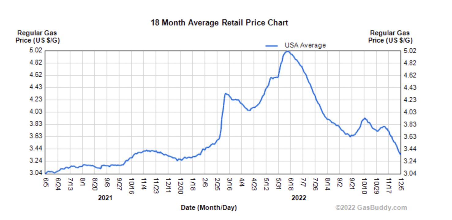
You know, if that last downward leg had happened a few weeks earlier, Ds would still control the House
Joseph P.
boosted

Matrix Vesicle Plasma Cell Membrane Glycoprotein-1
Regulates Mineralization by Murine Osteoblastic
MC3T3 Cells
authors : KRISTEN JOHNSON,1 ALLISON MOFFA,1 YING CHEN,1 KENNETH PRITZKER,2
JAMES GODING,3 and ROBERT TERKELTAUB
hey there folks, if you like this or know someone that might appreciate this stuff please drop me a toot and let me know : just trying to connect with folks.
ATP-Driven Molecular Chaperone Machines
#Molecular #chaperones are proteins that help maintain the balance of proteins in the #cell, which is essential for the cell to stay alive. Chaperones are always present in the cell, but they can also be activated in response to #stress. They interact with proteins that are not folded correctly, preventing them from clumping together and helping them to fold correctly. Chaperones don't usually interact with proteins that are already folded correctly. They use #energy from #ATP binding and/or hydrolysis to help with #folding and unfolding proteins. Because chaperones are involved in keeping protein balance, they are linked to diseases caused by #protein misfolding, such as #neurodegeneration and #cancer. Therefore, understanding how chaperones work is important for understanding and treating these diseases.
The Hsp70 system is a group of proteins that are found in #bacteria, #eukaryotic cells, and some #archaea. They are responsible for binding to unfolded or partially unfolded proteins to prevent them from aggregating and to help them fold correctly. Hsp70 proteins are made up of two parts: a 44 kDa N-terminal ATPase domain and a 28 kDa substrate binding domain with a C-terminal lid subdomain. The #ATPase domain helps the protein bind and release substrates, while the substrate-binding domain binds to extended #polypeptide chains. Hsp40 proteins, which are also known as J-domain proteins, act as co-chaperones to Hsp70 and help recruit substrates and stimulate the ATPase activity of Hsp70. Hsp40s can also direct Hsp70 to specialized functions and sub-cellular regions.
Hsp90 is a type of molecular chaperone that helps proteins fold correctly. It is made up of three conserved domains: the ATP binding N-terminal domain, the middle domain, and the C-terminal dimerization domain. Hsp90 works by binding to proteins that need to be folded correctly and preventing them from aggregating in an ATP-dependent manner. It also interacts with other proteins, called co-chaperones, which help regulate its ATPase cycle and determine which proteins it binds to. Hsp90 can also act as a buffer for genetic variation by rescuing mutated proteins with altered properties.
The different functions of the Hsp100/Clp proteins.
These proteins contain one or two conserved ATPases Associated with various cellular Activities (AAA1) domains and can act as #unfoldases or #disaggregases. Unfoldases help to unfold proteins and deliver them to a ring #protease, while disaggregases have the unique ability to recover proteins from both amorphous and #amyloid aggregates. The main difference between the two is the presence of a coiled-coil insertion in the first AAA1 domain in the disaggregases. The Hsp100 proteins usually form hexamers which hydrolyze ATP in either a sequential/random or a concerted manner. #Crystal structures have been determined of monomeric forms of several Hsp100 proteins, and of the hexamer forms of HslU, ClpX, and ClpC unfoldases. Hexameric forms of various Hsp100’s have been observed at intermediate resolutions by cryo-EM. These structures suggest a typical AAA1 packing arrangement for the unfoldases and an expanded conformation for the Hsp104 disaggregase. The central channels of the Hsp100s are lined by tyrosine residues, located on mobile loops, which bind substrates non-specifically. It is thought that rotations of the AAA1 domains provide the force to unfold the bound substrate and pull it through the channel. Disaggregation and unfolding functions are coupled and regulated via an interaction between the Hsp70 nucleotide-binding domain and the coiled-coil insertion. Recent biochemical and structural data suggest that it is docked on the outside surface of the AAA1 ring. Hsp100/Clp proteins are proteins that have one or two conserved ATPases Associated with various cellular Activities (AAA1) domains. These proteins can act in two different ways: as unfoldases or disaggregases. Unfoldases help to unfold proteins and deliver them to a ring protease, while disaggregases have the ability to recover proteins from both amorphous and amyloid aggregates. The main difference between the two is the presence of a coiled-coil insertion in the first AAA1 domain in the disaggregases. The Hsp100 proteins usually form hexamers which hydrolyze ATP in either a sequential/random or a concerted manner. Structures of these proteins have been determined, which suggest a typical AAA1 packing arrangement for the unfoldases and an expanded conformation for the Hsp104 disaggregase. The central channels of the Hsp100s are lined by tyrosine residues, which bind substrates non-specifically. It is thought that rotations of the AAA1 domains provide the force to unfold the bound substrate and pull it through the channel. Disaggregation and unfolding functions are coupled and regulated via an interaction between the Hsp70 nucleotide-binding domain and the coiled-coil insertion, which is docked on the outside surface of the AAA1 ring.
GroEL is a molecular chaperone machine that binds to proteins to prevent them from aggregating. It is estimated that GroEL binds to around 10% of the proteins in E. coli. The binding site is hydrophobic in character and contains essential hydrophobic residues that line the cavity-facing surface of the apical domain. If one of these residues is changed from hydrophobic to hydrophilic, the binding is abolished. Studies have shown that multiple binding sites act together as a continuous hydrophobic binding surface. It has also been shown that proteins stably bound to #GroEL are unstructured and that binding of non-native proteins to GroEL can be associated with unfolding. X-ray crystallographic studies have revealed structures of extended or helical peptides bound in the groove formed by helices H and I via hydrophobic interactions. Cryo-EM has also been used to probe the structure of non-native proteins bound to GroEL, which showed that the substrates were bound to helices H and I, with substrate density protruding from the GroEL ring. There is an upper limit, around 60 kDa, to the size of substrate that can fit inside the folding chamber. In summary, GroEL is a molecular chaperone machine that binds to proteins to prevent them from aggregating. It has a hydrophobic binding site that contains essential hydrophobic residues. Studies have shown that multiple #binding sites act together as a continuous hydrophobic binding surface and that proteins stably bound to GroEL are unstructured. X-ray crystallographic and cryo-EM studies have revealed structures of extended or helical peptides bound in the groove formed by helices H and I via hydrophobic interactions. The upper limit to the size of substrate that can fit inside the folding chamber is around 60 kDa.
Structural, biochemical, and biophysical studies have shown how proteins interact with GroEL, a protein-folding machine, and how ATP (a molecule that provides energy for many processes in cells) induces changes in GroEL's shape that allow it to switch between binding to proteins and folding them. Mutational analysis and cryo-EM studies (a type of imaging technique) have revealed that proteins primarily bind to a specific part of GroEL, and that multiple parts of GroEL bind to the protein at the same time. This binding causes the parts of GroEL to extend and expand, which helps to unfold the protein. Additionally, the protein's binding to GroEL provides a mechanical load on GroEL, which helps to further unfold the protein. Finally, when ATP is added, the parts of GroEL rotate, which removes the binding sites from the inside of the chamber and traps the protein in the chamber, which is now capped by GroES (another protein-folding machine). Group 2 #chaperonins are similar to GroEL, but have a slightly different structure. They are found in both eukaryotes (organisms with a nucleus, like humans) and archaea (a type of single-celled organism). They form back-to-back rings and have a high degree of sequence identity/similarity to GroEL. The main difference is an extension in the part of GroEL that forms the lid of the folding chamber. This extension helps to further unfold the protein.
The structural similarities between the Group 2 chaperonins and GroEL, both essential for folding proteins. Group 2 chaperonins have 8- or 9-fold symmetry, meaning that they form back-to-back rings with the same domain structure and a high degree of sequence identity/similarity to GroEL. The main difference between the two is an extension in the helix H equivalent in the apical domain of the Group 2 chaperonin, which removes the need for a GroES co-chaperone. Structural studies of the Group 2 chaperonins in different nucleotide bound states have revealed open, substrate binding and closed, substrate folding conformations similar to GroEL. These conformations involve a large clockwise twist of the apical domains and an inward tilt of the whole subunit, which brings the catalytic Asp in the intermediate domain close to the ATP binding site and closes the folding chamber. The ring expansion/contraction of Group 2 chaperonins is facilitated by the 1:1 nature of their inter-ring interface, allowing the equatorial domains to move more freely than in GroEL.
ATP-driven chaperones are proteins that help other proteins maintain their structure and function. They do this by binding, unfolding, refolding, and disaggregating proteins that are not in their native state. The Hsp70 system uses ATP binding and hydrolysis to regulate the binding and release of substrates. It is also regulated by co-chaperones. Hsp90 uses its ATPase cycle to induce multiple conformations that bind and stabilize or help mature the substrate proteins. It is also regulated by co-chaperones. The Hsp100s use ATP to unfold, thread, and disaggregate substrate proteins. In ATP-dependent proteolysis, the unfoldase is connected to a protease which breaks down the unfolded substrate proteins. The disaggregases, in combination with the Hsp70 system, use ATP-induced conformational changes to disaggregate and unfold substrate proteins. GroEL-GroES uses ATP binding to induce conformational changes to convert from a substrate binding to a substrate folding complex. It may also use the ATP-induced conformational changes to force the substrate to unfold. GroES binding then ejects the substrate from the binding surface, giving it a chance to fold in isolation during the slow ATP hydrolysis step. The archaeal Group 2 chaperonins use ATP-induced domain rotations that are similar to GroEL, but the conformational changes are not in the same order. The eukaryotic cytosol Group 2 chaperonin is similar to the archaeal system, but its ATPase cycle and action appear to be more complex and specific to the substrate.
In conclusion, ATP-driven chaperones are proteins that help other proteins maintain their structure and function by binding, unfolding, refolding, and disaggregating proteins that are not in their native state. Structural studies of GroEL and Group 2 chaperonins have revealed ATP-induced conformational changes that bind, unfold, and refold proteins in the ATP-dependent folding process. GroEL uses ATP binding to induce a conformational change that switches between the substrate binding and substrate folding states. The archaeal Group 2 chaperonins have a similar mechanism, but with a different order of conformational changes. The eukaryotic cytosol Group 2 chaperonin is more complex, indicating different substrate-specific functions. In general, ATP-driven chaperones help to ensure the proper folding of proteins and support the lifecycle of proteins.
If you like this post
👇
https://qoto.org/@tonic/109460888574379368
it was written using #Lex #ai and the game was in GPT-3
here's an invite to http://Lex.Page https://lex.page/invites/QCNQGOFT
Just comment or DM me for invites if it runs out 🚀
So i’m wholly convinced #AI has a pretty huge part to play in the future of education. Something like #ChatGPT let’s you have a didactic conversation about a lot of surface level topics. But beyond that you can give it personality by for example #gamify-ing the output to improve interest, for example in kids.
AI could help teachers in preparing materials and #grading, reducing the amount of time spilling out of their day.
I see AI as the logical next step in improving the #education system, by making it more efficient and productive while also providing a way to personalize #learning. AI can be used to create personalized learning paths, tailor-made to the style of learning of each #student, making sure they continue to nurture every one of their interests and passions. AI can also detect symptoms of #burnout or disengagement, and suggest activities to smooth their #journey.
For Example a student might be interested in a particular topic , but their parents not so much or their situation makes it hard to connect with #mentor s , #teacher s, role models, well with something like this they can really enjoy educating themselves on a given subject matter.
within a matter of seconds the AI dreamed up a text based #game to teach folks about #cell #metabolism.
jos****@****.***
create a text based decision game to help me learn about cell metabolism
Sure, I can help you create a text-based decision game to help you learn about cell metabolism. Here's a basic outline for the game:
Introduction: The game begins by introducing the player to the concept of cell metabolism and explaining the main objective of the game, which is to help a cell maintain its metabolic balance.
Decision-making: The player is presented with a series of choices that affect the cell's metabolic processes. For example, the player might be asked whether to increase or decrease the cell's production of a particular enzyme, or whether to activate a certain metabolic pathway.
Consequences: Each decision the player makes has consequences for the cell's metabolism. For example, increasing enzyme production might increase the cell's energy production, but it might also increase the cell's demand for oxygen. The player must weigh the pros and cons of each decision and decide which course of action to take.
Feedback: After each decision, the game provides feedback on the cell's metabolic status. This can include information on the cell's energy levels, oxygen levels, and other metrics that reflect the cell's overall health.
Repeat: The game continues in this manner, with the player making decisions and receiving feedback, until the cell's metabolism is in balance. At that point, the game is complete.
I hope this helps! Let me know if you have any other questions.
jos****@****.***
that sounds fun ! let's play the game I have 100 ATP points, let's use ATP to help balance the cell metabolism
Great! To start the game, you have 100 ATP points to use to help balance the cell's metabolism. In each round of the game, you will be presented with a decision that affects the cell's metabolism. For example, you might be asked whether to increase or decrease the production of a particular enzyme, or whether to activate a certain metabolic pathway.
Each decision you make will have consequences for the cell's metabolism. For example, increasing enzyme production might increase the cell's energy production, but it might also increase the cell's demand for oxygen. You must weigh the pros and cons of each decision and decide which course of action to take.
After each decision, the game will provide feedback on the cell's metabolic status. This can include information on the cell's energy levels, oxygen levels, and other metrics that reflect the cell's overall health.
The game continues in this manner, with you making decisions and receiving feedback, until the cell's metabolism is in balance. At that point, the game is complete.
Good luck! Let me know if you have any questions as you play the game.
jos****@****.***
I decide to spend some ATP to produce NAD+
Spending ATP to produce NAD+ is a good decision, as NAD+ is an important cofactor that is involved in many cellular processes, including energy metabolism. Producing NAD+ can help the cell maintain its metabolic balance and support its overall health.
However, it's important to consider the potential costs and benefits of this decision. For example, increasing NAD+ production might require the cell to use more ATP, which could deplete its energy stores. It's also possible that increasing NAD+ production could affect other metabolic processes in the cell, such as oxygen levels or the production of other enzymes.
Therefore, it's important to carefully consider the potential consequences of your decisions and weigh the pros and cons before making a decision. Good luck! Let me know if you have any other questions.
Joseph P.
boosted

RT @_JosephWatson@twitter.com
Today we’re sharing a deep-learning method for protein design called RoseTTAFold Diffusion. With minimal input, it turns prompts (“create a molecule that binds X”) into new proteins that fold and function in the lab. We’ve tested 100s already.
PDF here: https://www.bakerlab.org/2022/11/30/diffusion-model-for-protein-design/
🐦🔗: https://twitter.com/_JosephWatson/status/1598409457855496192
Joseph P.
boosted

I listened to this bite-sized pod interview of Adam Grant who said “these days we are more interested in proving ourselves than improving ourselves”. I really felt this because I have (and I know many of my colleagues also have) very slim margins for errors or setbacks. I’m terrified of awful reviews (I’ve had m.a.n.y.) and I spend way more time making my work perfect than on learning and broadening my skills and thinking. The last time I lived by this attitude, I was not a PI.
Joseph P.
boosted

One thing I've noticed after years of exercise... I am willing to put in the effort neccesary to do the carb loading needed to win marathons... its the running part im not to keen on...
Joseph P.
boosted
Adenosine, a #biochemical that is found in cells and is essential for many processes. Adenosine is made up of an adenine nitrogenous base linked to a #ribose sugar #molecule. It is involved in the #biosynthesis and #replication of #DNA and #RNA, and is also a component of an #enzyme called S-adenosyl-L-methionine (SAM) and cyano-cobalamin (vitamin B-12). #Adenosine was once thought to only modify #cardiovascular #physiology, but now it is known to have many other effects on physiology.
Joseph P.
boosted

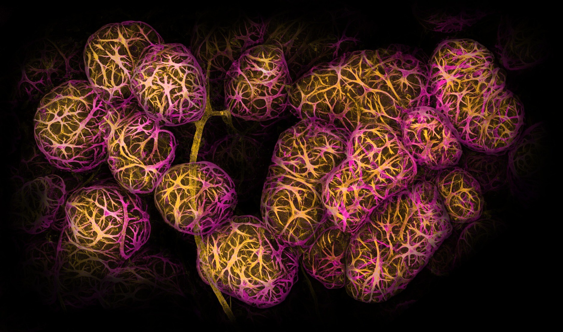
Calling all Mastodonians!
Let's make #ScienceSunday a trending hashtag every weekend. Post anything related to science: a cool image, research article, or scientific observation. Look to the stars and skies, to nature, or even the food you cook for inspiration.
Tag your post and boost other tagged posts generously.
Here's a prize-winning image by Dr. Caleb Dawson from 2022 Nikon Small World competition. Can you guess what it is?
Answer in the image description.
Joseph P.
boosted
Just a reminder, as I get this a lot, but joining the United Federation of Instances doesnt require **any** time contribution of the part of its admins or instances. Any voting is totally optional to participate. As long as your instance follows our code of ethics you can join, nothing else required other than to federate to other UFoI instance.
DM me if you want to talk more, are are at over 200,000 members and will be open to more over the next week or so before membership will become harder and more formalized.
all intances welcome, from single user to large instances.
I'm playing my part to support #biomanufacturing #biofabrication #biofoundry(ies) and bear witness to the birth of an industry. Working with 2 novel compounds to grow bone, cartilage & other types of cells. turns out it also works for growing hearts @organamet
Joined Nov 2022
